The Kidney

The kidney is commonly involved by various subtypes of systemic amyloidoses with light chain amyloidosis (AL) being the most frequent cause of renal amyloidosis followed by amyloidosis A (AA), leucocyte chemotactic factor 2 (ALECT2) then fibrinogen (AFib). (see section on Nomenclature)

However, in certain non-Anglo Saxon ethnic groups the non-AL types of AA and ALECT2 amyloidosis are more common.

There can be many pitfalls in the typing of amyloidosis and the correct typing of renal amyloid is crucial to avoid unnecessary toxicity from misdirected AL chemotherapy.

Each type of amyloidosis is a distinct disorder with different prognoses, renal recurrence risk, extrarenal organ involvement and management.

Proteinuria and renal impairment are the most common manifestations of renal amyloidosis.

A biopsy is required to identify renal amyloidosis. However, in confirmed AL amyloidosis diagnosed by biopsy of other organs, renal amyloidosis can be assumed if there are no other causes for a coincident proteinuria.

Typing the renal amyloidosis requires clinico-pathologic assessment with monoclonal gammopathy testing to screen for AL, organ staging and amyloid protein typing.

In some cases, specialised testing may be required with;

  • Amyloid protein typing by mass spectrometry
  • Genetic screening for amyloidogenic mutations

Familiarity with the demographic, clinical, renal and histopathological characteristics of renal amyloidosis is required to accurately diagnose and type renal amyloidosis.

The AAN’s clinics and laboratory services were established to assist in the typing and management of amyloidosis and welcome all referrals.

Amyloidosis and the Kidney

Epidemiology

KEY POINT

  • Amyloidosis is an infrequent cause of nephrotic syndrome and end-stage kidney disease
  • Kidney involvement is common in several types of systemic amyloidosis
  • AL is the most common amyloidosis affecting the kidneys causing at least 80% of renal cases, followed by AA, ALECT2 then AFib amyloidosis

Amyloidosis is an infrequent cause of nephrotic syndrome and end-stage kidney disease

Kidney involvement is common in several types of systemic amyloidosis

AL is the most common amyloidosis affecting the kidneys causing at least 80% of renal cases, followed by AA, ALECT2 then AFib amyloidosis

Amyloidosis is an infrequent cause of nephrotic syndrome with an incidence rate that increases with age.1

AL amyloidosis is the most common type of renal amyloidosis accounting for >80% of cases with local data 2 showing a similar distribution of renal amyloid types as observed at the Mayo Clinic 3 (Table 1):

Table 1. Types of Renal Amyloidosis

Amyloidosis Type 

AAN (VTAS) Clinic (N=38)

Mayo Clinic (N=474)

Immunoglobulin derived i.e. AL/AH

31 (82%)

407 (85.9%)

Non-AL

7 (18%)

67 (14%)

AA

ALECT2

AFib

4 (10%)

2 (5%)

1 (3%)

33 (7%)

13 (3%)

6 (1%)

Others

0 (0%)

15 (3%)

Unclassified

AApo

Combined*

11 (2.3%)

3 (0.6%)

1 (0.2%)


Key: AA = Serum amyloid A, AFib = Fibrinogen alpha, AH=Immunoglobulin heavy chain, ALECT2= Leucocyte chemotactic factor-2, AL=Immunoglobulin Light chain, AApo=Apolipoprotein A variants (n=1 for each of AApo AI, II, IV), VTAS=Victorian and Tasmanian Amyloidosis Service
* The one combined case was that of AA amyloidosis and Ig heavy and light chain amyloidosis

However, in certain non-Anglo Saxon ethnic groups AL is not the most common amyloidosis type eg for Egyptian patients both AA (48%) and ALECT2 (31%) are more common than AL (20%). 4

The kidney is commonly involved by systemic amyloidosis but there is a wide range in frequency of renal disease according to the amyloidosis type. The rate of renal amyloidosis in:

  • ALECT2 and AFib: is almost universal 4
  • AA: is ≈ 97% 5
  • AL: is ≈ 70% 6
  • ATTR: extremely rare as it is not seen in ATTRwt (the most common type of transthyretin amyloidosis) and is restricted to some ATTRv gene mutations 7

Amyloidosis is an uncommon cause of end-stage kidney disease (ESKD) accounting for approximately 0.8% of patients requiring renal replacement therapy (RRT) in Australia and New Zealand. 8,9 The risk of progression to ESKD is mainly dependent on the amyloidosis type and the availability of disease specific therapy.
Kidney transplantation is rarely performed due the difficulty in ensuring graft survival in the most common type of renal amyloidosis (AL). For instance, less than 1% of ESKD patients with AL amyloidosis received a kidney transplant in the United Kingdom between 1984 and 2009. 10

However, other types of renal amyloidosis are more suitable for the option of renal transplantation as;

  • Amyloid recurrence in the renal allograft and/or graft failure may take more than several years to occur eg as in AFib and ALECT2 amyloidosis
  • Durable disease modifying therapies may be available eg as in AA

Anatomical Pathology

KEY POINT

  • Renal amyloidosis is diagnosed on biopsy using the Congo Red stain followed by examination under polarised light
  • Characteristic amyloid distribution patterns in the kidney can provide useful clues to the amyloid type
  • Immunohistochemistry (IHC) amyloid typing stains are routinely performed but it is important to note that their positive predictive value is only ≈ 60% and that stains for non-AL/AA renal amyloid types are not readily available
  • Laser microdissection of the amyloid deposit followed by mass spectrometry of the constituent protein is used when the amyloid type remains uncertain

Renal amyloid under light microscopy is identified by glomerular deposits of amorphous hyaline material which is Congo Red stain positive displaying apple green birefringence and dichroism effects under polarised light.

In the most common types of renal amyloidosis (AL and AA) the amyloid is deposited predominantly in the glomeruli and vessels and less commonly in the interstitium.

Characteristic patterns of amyloid deposition can be useful in typing other renal types of amyloid however these features can overlap and are not pathognomonic. The characteristic glomerular deposition patterns can be summarised as follows:

  • AFib: Vascular
  • ALECT2: Cortical interstitial
  • AApo: Medullary interstitial

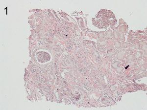
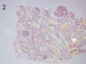
Photos 1 and 2. ALECT2. The cortical interstitial deposition pattern of Congo Red staining (1) of ALect2 renal
amyloid under light microscopy and polarized light (2). Photos courtesy of Dr Shannon Bahamehr Fadaee.


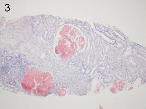
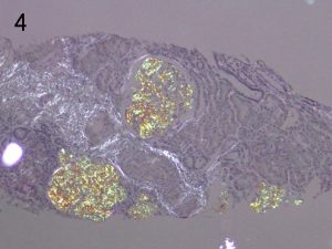
Photos 3 and 4. AFib. The glomerular deposition pattern of Congo Red staining (3) of AFib renal
amyloid under light microscopy and polarized light (4). Photos courtesy of Dr Shannon Bahamehr Fadaee.


On electron microscopy the amyloid is characterised by randomly arranged, small organised fibrils, with a diameter of 8-15 nm. The alternative diagnosis of fibrillary glomerulonephritis can be differentiated by;

  • A lack of positive Congo Red staining
  • Larger fibrils (12-24 nm) in diameter although there is an overlap
  • Positive DNAJB9 immunohistochemistry (IHC) staining which has 98% sensitivity and >99% specificity for fibrillary glomerulonephritis 11

IHC amyloid typing stains should always be performed. However, it is important to acknowledge that false positives and false negatives occur due to intrinsic limitations in the antisera used and that the positive predictive value of these stains is only in the order of ≈ 60%. 12,13 See anatomical pathology section The basic IHC typing panel for renal amyloid includes antisera against the following amyloid fibril proteins:

  • Lambda and Kappa light chain
  • AA

Immunohistochemical amyloid typing stains for rarer amyloid proteins are not readily available in Australia eg the ALECT2 and AFIb stains. And non-AL nan-AA cases are typically referred to reference labs for further typing by second tier IHC stains and if required mass spectrometry. See special tests section

Clinical Presentation

KEY POINT

  • Renal amyloidosis usually presents with proteinuria and/or impaired kidney function
  • The amyloid type and amyloid burden and its distribution in the kidney influence the clinical presentation
  • The most common type of renal amyloidosis is AL amyloidosis and this typically presents with multiorgan disease
  • The next most common types AA, ALECT2 and AFib are usually associated with isolated kidney disease

The usual presenting features of renal amyloidosis are proteinuria and/or impaired kidney function. However, each type of renal amyloidosis can be considered as separate disorders with different levels of proteinuria, rates of progression to ESKD, extra-renal manifestations and therapy.

The proteinuria may be mild through to nephrotic in range. ALECT 2 often presents with minimal proteinuria (as the amyloid is predominantly interstitial) and typically remains subnephrotic even when there is progression to ESKD. The proteinuria of AL and AA is of a glomerular pattern with the capacity to develop nephrotic range proteinuria.

The kidney dysfunction can range from minimal to end-stage kidney disease (ESKD) requiring dialysis. The rate of progression to ESKD also varies and is slower in ALECT2 than in AL with ALECT2 associated with a mean eGFR loss of only 4.2 mL/min/yr. 15

AL is commonly associated with multi-organ amyloidosis whereas the other types of are more likely to present with isolated renal disease. Of important prognostic significance, the heart is commonly involved in AL but only rarely in AA (≈2%) and is not described as yet in ALECT2.

There are other clinical features that can assist in the typing of the renal amyloid;

  • ALECT 2 is typically seen in non-Anglo Saxon ethnic groups, particularly patients of Egyptian, Hispanic, Indian and Pakistani background (and it has also been recently described in the Chinese population) 16
  • AA amyloidosis is classically associated with disorders causing uncontrolled inflammation eg an active autoinflammatory disorder such as rheumatoid arthritis, chronic infection or inflammatory malignancies (typically renal cell cancer.) However, it has become increasingly common for the inflammation to be of uncertain origin, a phenomenon which may be partly related to obesity and its associated low-grade chronic inflammation.

For hereditary amyloidosis there may not be any clear family history eg there is no family history in >50% AFib despite an autosomal dominant inheritance pattern and this is partly due to the incomplete penetrance pattern of the hereditary amyloidoses. 17

The Workup and Typing of Renal Amyloidosis

KEY POINT

  • After a kidney biopsy has identified amyloidosis the basic workup starts with:
    • Screening for AL with monoclonal gammopathy testing
    • and
    • Organ staging
  • Renal involvement may confound the interpretation of some of the basic workup tests
  • Monoclonal gammopathy screening is an essential part of the assessment of renal amyloidosis, but caution is required with interpretation of the serum free light chains (SFLC) in the setting of impaired kidney function

After a kidney biopsy has identified amyloidosis the basic workup starts with;

  • Screening for AL by assessing for a monoclonal gammopathy
  • and
  • Organ staging

The components of the basic workup (Diagram 1) are discussed in more detail elsewhere.

Diagram 1. The Basic Workup after Identification of Renal Amyloidosis

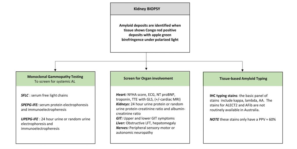
Key: AA- = Amyloid A, AFib = Fibrinogen amyloid, amyloid ALECT2 = Leucocyte chemotactic factor 2 amyloid, ECG = electrocardiogram, GIT= gastrointestinal tract, GLS= global longitudinal strain, LFT=liver function tests, MRI= magnetic resonance imaging, NT proBNP= N-terminal pro brain natriuretic peptide, PPV= positive predictive value, TTE= transthoracic echocardiogram

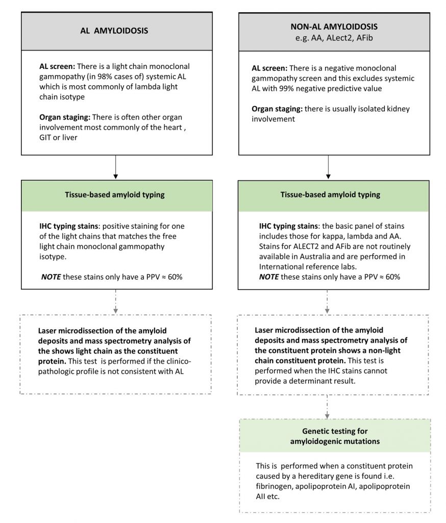
A summary of the different diagnostic findings for AL vs non-AL renal amyloidosis is diagrammatically presented (Diagram 2).

Diagram 2. The Test findings of AL vs non-AL Amyloidosis

AA= Amyloid A, ATTR= transthyretin amyloid, ECG = electrocardiogram, GIT= gastrointestinal tract , GLS= global longitudinal strain, IHC= immunohistochemical, Ix= investigation, LFT=liver function tests, MGUS= monoclonal gammopathy of uncertain significance, MRI= magnetic resonance imaging, NT proBNP= N-terminal pro brain natriuretic peptide, PPV= positive predictive value, TTE= transthoracic echocardiogram

Renal failure can confound the interpretation of many of the basic workup tests and the issues are discussed below.

Monoclonal Gammopathy Tests

Testing for the monoclonal gammopathy associated with AL requires all three of;

  • Serum Free Light Chains (SFLC)
  • Serum Protein Electrophoresis and immunofixation (SPEP + IFE)
  • Urine Protein Electrophoresis and immunofixation (UPEP + IFE)

98% of AL amyloidosis cases have a light chain monoclonal gammopathy whereas only 50% have an associated paraprotein. However, the presence of a monoclonal gammopathy does not always indicate AL renal amyloidosis as monoclonal gammopathy of uncertain significance (MGUS) particularly those with just a paraprotein is common. 18

Positive identification of an amyloidogenic light chain is currently defined as an elevated kappa or lambda light chain together with an altered kappa to lambda ratio; 13

  • The median monoclonal light chain level at presentation is only 100mg/L
  • In ≈80% the monoclonal light chain is a lambda light chain 19

Interpretation of the serum free kappa:lambda light chain ratio requires caution in patients with impaired kidney function. The normal range for the kappa:lambda ratio is 0.26 to 1.65. However, the kappa:lambda ratio may increase to 3.1 in those with ESKD (requiring dialysis) due to the reduced clearance of kappa light chains over lambda light chains. 20 This can lead to a:

  • Misdiagnosis of kappa monoclonal gammopathy in cases with a kappa/lambda ratio of between 1.65 and 3.0 (false positive)
  • Missed identification of a lambda monoclonal gammopathy in cases of lambda AL amyloidosis with impaired kidney function and a low-normal kappa:lambda ratio or inappropriately increased lambda FLC (false negative)

Organ Staging

The organ staging of the heart can be confounded by renal failure. This is a common issue in AL amyloidosis where both kidney and heart disease can occur.

The NT proBNP and troponin are sensitive screening biomarkers for cardiac amyloidosis but can also become elevated by impaired kidney function

Ventricular thickening on TTE can occur in those with ESKD on haemodialysis

Cardiac MRI with gadolinium is contraindicated in patients with stage 4 or 5 CKD with eGFR<30mL/min/1,73m²

  • However, cardiac MRI without gadolinium can be used in such patients where “Native” (or non-contrast) T1-myocardial mapping techniques indicate the presence of cardiac amyloidosis by showing extracellular volume expansion with significantly increased native T1 times see Heart Investigation Findings / Non-invasive Imaging

Specialised Typing Tests

Further tests may be required to type the renal amyloid and these include:

1. Proteomic analysis where laser microdissection of the amyloid deposit from the paraffin embedded tissue block is followed by trypsin digestion then tandem mass spectrometry analysis of the constituent amyloid protein. 21 This is most commonly required in isolated renal disease occurring in the context of;

  • The lack of a monoclonal gammopathy
  • Uninterpretable SLFC in the setting of severe CKD
  • Possible cases of AA where there are inconsistent AA stain and clinical findings

Mass spectrometry is then required and usually diagnoses the following;

  • ALECT2
  • Hereditary amyloidoses eg AFib and AApo
  • AA (usually in a patient without a clear inflammatory disorder)
  • Rarer amyloidoses eg heavy chain amyloid

2. Genetic testing eg for AFib

Genetic testing is performed after mass spectrometry has identified an amyloid precursor protein with a hereditary genetic cause. See Specialised Tests

Clinicopathologic profiles

There are helpful clinico-pathologic characteristics for the different types of renal amyloid but they should not be used to definitively type the amyloidosis (Table 2).

Table 2: Clinical and histopathological characteristics of the most common types of renal amyloidosis

Type Renal and clinical  presentation Renal compartment involved Supporting/ confirmatory tests
AL Proteinuria, nephrotic syndrome, impaired kidney function

+/- cardiac amyloid

Glomerular + Vascular > Interstitium Light chain monoclonal gammopathy (usually lambda)

Light chain (usually lambda) restriction on IHC staining. Note: Light chain IHC stains only have a PPV of approximately 60%

AA Proteinuria, nephrotic syndrome, impaired kidney function

Inflammatory conditions i.e. rheumatoid arthritis, chronic infection, renal cell cancer, obesity

Glomerular + Vascular > Interstitium AA on IHC staining

Raised Serum Amyloid A and/or CRP

ALECT2 Often minimal proteinuria, impaired kidney function

Median age at presentation of 62-68 years

Specific for certain non-Anglo Saxon ethnic groups (Egyptians, Hispanics, Indians and Pakistani)

Interstitium +++ IHC staining for LECT2 is reliable but not available in Australia

Mass spectrometry showing LECT 2 protein as the constituent amyloid protein is often required

AFib Proteinuria, impaired kidney function

Median age at presentation of 58 years

No family history in >50%

Glomerular +++ IHC staining for Fibrinogen (not easily available in Australia)

Mass spectrometry showing fibrinogen protein as the constituent amyloid protein is often required

Genetic testing of the Fibrinogen exon of interest

Key; AA= serum Amyloid A amyloid, AL= light chain amyloid, ALECT 2= leucocyte chemotactic factor amyloid, CRP= C reactive protein, IHC= immunohistochemistry, PPV= positive predictive value

Prognosis

KEY POINT

  • The patient and renal prognosis varies according to the amyloidosis type
  • Amyloid specific therapy is available for AL and AA
  • Early treatment is crucial as deep suppression of amyloid synthesis is associated with organ responses and improved renal and patient survival outcomes
  • Renal amyloid clearance can occur after disease modifying treatment, but achieving a renal response often takes more than 6 months after stopping the amyloid forming process

The patient’s overall survival and renal prognosis varies according to the;

  • Type of amyloidosis
  • Degree of renal involvement
  • Availability and efficacy of therapy

The renal prognosis can improve with treatment as renal amyloid clearance can occur by the proteolytic breakdown of amyloid fibrils by phagocytic cells if amyloid production is stopped (or markedly reduced) by disease modifying treatment. This process is slow and achieving a renal response often takes more than 6 months after an effective reduction in amyloid synthesis. Disease modifying therapy and hence the chance of renal recovery exists for both AL and AA amyloidosis.

AL

Patient survival is poorest for AL amyloidosis as multiple organs are often affected. The main predictor of survival in AL is the presence and degree of cardiac involvement however the requirement for dialysis can also impact on survival.

The haematologic response reflects the of depth of the reduction in production of amyloidogenic light chains. Deep haematologic responses (of a “very good partial response” or better) are associated with median OS rates of > ≈ 80% at 2 years 22 and can even prolong the survival of those with advanced cardiac disease. 23 See Response tab of AL Amyloidosis

A renal prognostic staging system has been developed for newly diagnosed AL using the cut-offs of an eGFR 50mL.min/1.73m2 and a 24 hour proteinuria level of 5g/day at baseline. 24,25 (Table 3)

Table 3: AL Renal Prognosis. Based on Dispenzieri A et al “Renal Risk and Response in Amyloidosis”. 25

Renal Stage Baseline eGFR (mL/min/1.73m²) & proteinuria

2-year risk of dialysis (%)

Annual risk thereafter (%)

Stage I eGFR ≥50

AND

Proteinuria <5 g/day

0 – 3 0-1
Stage II EITHER
eGFR <50

OR

Proteinuria ≥5 g/day

11 – 25 <5
Stage III eGFR <50

AND

Proteinuria ≥5 g/day

60 – 75 0 – 15

With a haematologic response (HR), an organ response can occur and ≈ 65% of those who develop a HR go on to achieve a renal response. 25 The depth and speed of the haematologic response also affect the renal response rate. 26,27

Table 4: The depth and speed of haematological response predicts renal response in AL amyloidosis 26

% dFLC reduction at 6 months Probability of Renal Response (%)
0-49 19.2
50-90 29.4
>90 89.1

Key: dFLC (difference in free light chains)= amyloidogenic free light chain minus the uninvolved free light chain

The criteria for a renal response in AL are a;

  • ≥30% decrease in proteinuria
  • or
  • decrease in proteinuria to below 0.5 g/day
    in the absence of

Renal progression which is defined as a ≥25% decrease in eGFR 28

The achievement of a renal response reflects amyloid proteolysis and predicts for dialysis independence. 24, 25 Proteolytic breakdown of amyloid fibrils is mediated by phagocytic cells and is a slow process. For an organ response to occur the rate of amyloid production needs to be slowed to below the rate of proteolysis.

AA

The median overall survival was found to be 133 months (after diagnosis) in the largest cohort analysis of AA amyloidosis patients. 29

Approximately 48% of patients develop ESKD and of these 53% present with ESKD at diagnosis and for the remainder, the median time from presentation to ESKD is 26 months. 5

As for AL, the renal prognosis can be improved by the early institution of effective directed treatment against the underlying inflammatory process. See Amyloidosis AA section

Patient survival and renal outcomes are correlated with the serum Amyloid A (SAA) level link to contacts>lab where the lower the SAA level the better the outcomes; 29

  • Amyloid deposits can regress in 60% of cases where the median SAA is reduced to <10 mg/L 29

It is ideal to maintain the SAA within the reference range and to aim for even the lower part of the reference range as incremental benefits in survival continue to be observed when the SAA is reduced below the upper limit of the reference range (of 6.4mg/L).

ALECT2

ALECT 2 amyloidosis was only discovered in 2008 and therefore little is known about this disorder. See “ALECT2 and Other”.  ALECT2 has a predilection for the kidneys and the liver although the latter is usually asymptomatic.

There is no specific therapy for ALECT2.

The data on renal prognosis is limited and based on small case series. There is a slow renal progression rate with a mean eGFR loss of 4.2 mL/min/year.17
The reported rate of progression to ESKD is wide ranging (25% to 46%) and probably reflects the lack of longterm epidemiologic data. The median time from diagnosis to ESKD is 8-16 months (range 0-73) and is dependent on the disease extent at diagnosis. 16,30

AFib

In a United Kingdom cohort of AFib patients, 62% developed ESKD by a median age of 60 years (range 36-82) and the median time from presentation to ESKD was 4.6 years.17

There is no specific therapy for AFib amyloidosis. See Hereditary Amyloidosis section>AFib

Supportive Care

The management of renal amyloidosis can be divided into disease modifying treatment and supportive care. Disease modifying therapy is described elsewhere on this website under each type of amyloidosis.

The supportive care of nephrotic syndrome is discussed in this section of the website and addresses;

  • Fluid retention
  • Thromboembolism
  • Hyperlipidaemia

Generic supportive therapies for nephrotic syndrome are not always generalisable to patients with renal amyloidosis.

Renal replacement therapy is discussed and divided into;

  • Dialysis
  • Kidney transplantation

Palliative care (without dialysis) should be considered for those with poor prognosis and quality of life.

Renal supportive care is challenging and requires a cautious approach with constant review and adjustment.

Nephrotic syndrome

Oedema

For oedema the usual supportive principles apply but the anti-proteinuric effects of angiotensin converting enzyme (ACE) inhibitors and angiotensin receptor blockers (ARB) remain unproven and prone to causing side effects particularly in AL patients.

The main measures to counteract the oedema caused by nephrotic syndrome are;

  • Salt and fluid restriction
  • Diuretics
    • Loop diuretics (eg frusemide) are the first-line therapeutic class of diuretics. In refractory cases, agents that react more distally in the nephron are considered depending on serum potassium and kidney function and include thiazide diuretics (e.g. hydrochlorothiazide) or potassium sparing diuretics (eg. spironolactone)
    • Although ACE inhibitors and ARBs are often used in other disorders that cause nephrotic syndrome for their anti-proteinuric effect, their utility remains unproven in amyloidosis. These agents may not be tolerated and should be used with caution with concomitant diuretics and in the presence of borderline blood pressure or hypotension
  • Salt-poor albumin infusions can be considered as an adjunct to allow effective diuresis in overloaded patients
  • Patient education of;
    • Factors that influence fluid status eg temperature/weather, medications, salt intake
    • Sodium and fluid monitoring and restriction
    • Monitoring with daily weighs

Thromboembolism

The risk of thromboembolism increases as the serum albumin decreases but the patient specific risk of bleeding should also be considered. The decision to commence anticoagulation should be individualised and the risk of venous thromboembolism balanced against the risk of bleeding.

The bleeding risk is increased when there is;

  • GIT amyloidosis
  • Acquired factor X deficiency (a rare and specific phenomenon of AL)
  • Treatment-related thrombocytopaenia (a potential issue with some AL therapies)

Important non-renal risk factors for thrombosis to consider are;

  • Prior history of venous thromboembolism
  • Use of drugs in the immunomodulatory therapeutic class used in AL (thalidomide, lenalidomide and pomalidomide)

In general;

  • Anticoagulation is not indicated if the serum albumin is >30 g/L
  • Consider anticoagulation if the bleeding risk is low and the serum albumin is <20 g/L

The choice of anticoagulation is between warfarin or low molecular weight heparin with the latter adjusted for GFR. There is limited data on the new oral anticoagulants (NOAC) in nephrotic syndrome. There have been case reports of breakthrough thrombosis whilst on NOAC therapy hypothesised to be due to the unsustained plasma levels of highly protein bound NOAC in the hypoalbuminaemic state. NOACS are only considered if other options are not tolerated or declined by patients.

Hyperlipidaemia

The long-term consequences of prolonged dyslipidaemia in nephrotic syndrome remains poorly understood. Adults with nephrotic syndrome have a markedly increased risk of both myocardial infarction and coronary death compared with that of healthy individuals. Unfortunately, there is very little data on the effects of statins on cardiovascular endpoints in nephrotic syndrome. 31 A meta-analysis that included four randomized controlled trials comparing statins versus placebo versus no treatment showed limited effectiveness of statins in reducing dyslipidaemia in nephrotic syndrome. 32

Despite the absence of clear guidelines regarding treatment, various strategies can be contemplated as statins and other lipid lowering therapies are well tolerated and may be useful.

Dialysis

In regards to dialysis modality, both haemodialysis and peritoneal dialysis are acceptable and the choice depends on the standard non-amyloid parameters as well as amyloidosis specific factors;

  • Haemodialysis may not be well tolerated with hypotension and/or cardiac amyloid
  • For peritoneal dialysis considerations are;
    • Increased risk of peritonitis if immunosuppressed
    • Possible worsening of hypoalbuminaemia
    • Less effective fluid removal if already oedematous

The standard indications for dialysis commencement apply if the patient is not an AL autologous stem cell transplant (auto-SCT) candidate.

For the auto-SCT candidate with near ESKD, it is generally recommended that;

  • Dialysis be commenced prior to ASCT
  • Haemodialysis be commenced via an arteriovenous fistula (AVF) with adequate anticipation time for the AVF to mature (>6 weeks)
  • A Permacath can be used but this is associated with a risk of line-related sepsis
  • Peritoneal dialysis should be avoided until adequate recovery of immunity post-ASCT due to the risk of peritonitis
  • If haemodialysis is the planned long-term dialysis modality avoidance of PICC lines, cannulation and venepuncture in the non-dominant arm is advised to preserve veins for future AVF creation

Kidney Transplantation

Historically, very few patients with amyloidosis (especially those with AL) have received kidney transplantation. However, treatment for AL amyloidosis has improved in the contemporary era leading to improved response rates and prognosis.

Medical suitability for kidney transplantation is be based on standard criteria as well as the following amyloidosis factors;

  • Type of amyloid
  • Prognosis
  • Treatment availability and response
  • Presence of extrarenal involvement
  • The risk of recurrence and premature transplant failure

For deceased donor kidney transplantation in Australia, candidates should have a low anticipated perioperative mortality and a reasonable estimated post-transplant patient and transplant survival as per “The Transplantation Society of Australia and New Zealand: Clinical Guidelines for Organ Transplantation from Deceased Donors”

According to the international “Kidney Disease Improving Global Outcomes (KDIGO) Clinical Practice Guideline on the Evaluation and Management of Candidates for Kidney Transplantation” candidates with;

  • AL amyloidosis should only be considered for transplantation if there is no or minimal extrarenal disease (particularly cardiac amyloidosis) and where a “potentially curative” treatment regimen has induced a stable remission
  • AA amyloidosis can be considered after adequate treatment of the underlying cause and in the absence of severe extrarenal organ involvement

Such guidelines adhere to the requirement for reasonable patient and transplant outcomes after renal transplantation.

AL

For AL amyloidosis, renal transplantation can be considered after achieving a sustained deep response in a patient with no or minimal cardiac involvement.

Kidney transplantation can be considered after achieving a complete remission (CR) or very good partial response (VPGR) for at least 12 months. Deep sustained responses are most often observed after autologous stem cell transplant (Auto-SCT).

Reported long term outcomes on renal transplantation outcomes in AL have shown; 33

  • 1 and 5-year median graft survival rates of 94% and 81% respectively
  • Improved median transplant survival in those with deep haematologic responses (10.4 versus 5.5 years)
  • Recurrent graft amyloidosis in 29% the graft after a median time from kidney transplantation of 3.7 years (range: 1.1-11.9) with transplant failure in 29%

An important issue with renal transplantation in AL is the chance of rejection from immunomodulatory drugs. There have been case reports of lenalidomide eliciting unintended immune activity against allografts in solid organ transplant recipients.34,35

AA

Kidney transplantation can be considered after adequate treatment of the underlying inflammatory cause and is associated with a low risk of recurrence in the transplant.

AA patients are generally good candidates as they usually only have renal disease with a low rate of extra-renal disease eg only ≈ 2% develop cardiac AA amyloidosis. See Amyloidosis AA

ALECT2

There is little reported experience with renal transplantation in ALECT2 however kidney transplantation can be considered if there is no other contraindication.

ALECT2 is a good prognosis amyloid group with slow progression and a lack of cardiac disease.

1 in 5 patients can develop amyloid graft recurrence but this does not seem to be associated with significant renal impairment and occurs after a relatively long time eg in one study, after a median follow-up of 20 months. 30

AFib

AFib is another good prognosis group where the heart is only rarely involvement.

Graft survival is excellent and after excluding early graft failures the 5 year and 10 year graft survival has been reported to be 100% and 68% respectively. 35

Combined liver-kidney transplant has historically been performed to restore kidney function and to stop the liver synthesis of AFib. This approach is no longer generally recommended due to significant perioperative risk but it is still sometimes considered in younger patients. 37,38